The Head of the Independent Research Group:
dr n. med. Tomasz Kolanowski
email: tomasz.kolanowski@igcz.poznan.pl
phone: +48 61 65 79 219
In order to push the human development, disease mechanisms and therapeutic strategies discoveries we need reliable, accessible yet physiologically relevant models. Laboratory animal–based research, although crucial for basic research, often show discrepancies between existing knowledge and in vivo reality in humans. This leads to number of clinical trial failures of leads (drugs) with otherwise good preclinical performance.
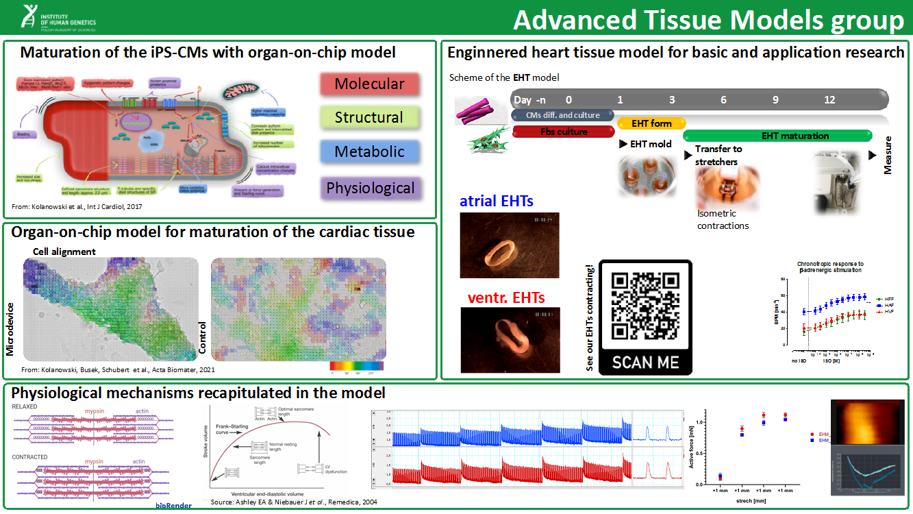
Our group has been founded in response to need of development new therapeutic models for basic discorevy research and drug screening purposes. We employ (a) simple (incl. organoids), (b) more advanced (lab-on-chip) and (c) macro-scale (engineered heart tissue, EHT) models in order to follow basic mechanisms of tissue formation and maturation. We apply our models to investigate not only molecular background of terminal cell differentiation and maturation but also micromechanics of the tissues to define causes of thus far idiopathic diseases and for pharmacological studies.
Currently we work with induced Pluripotent Stem Cell-derived cardiomyocytes (iPSC-CMs) in order to define subtype differentiation of the atrial and ventricular cardiomyocytes (aCMs vs vCMs). A exemplary FIBROdrive project employs Engineered Heart Tissue model – iPSC-CMs, cardiac fibroblasts and extracellular matrix component to define the external microenvironment needed to drive maturation of the CMs. Examples of conducted research can be found in the Figure 1.
In our research we employ a PANAKEIA platform that fuses super-resolution confocal and atomic force microscopy as well as provides electrophysiological studies by employing patch clamp technique for evaluation of advanced models (with organ-on-chip platform).